Veterinary Science: lameness in horses.
Unlike in humans, horse limbs have no bone connections with the torso (e.g. it has no clavicle), and the two sides are linked only and exclusively thanks to muscles. The shoulder blade, with its muscles and ligaments, is the only bone link with the torso.
To continue, between the scapula and the torso, a slipping movement is established that allows for the forward or backward movement of the limbs. The shoulder muscles are linked to the forearm muscles, which are divided into the following: extensors, flexors and adductors.
For the anatomical structure, they are specialised in the function of contraction and they allow for the activities of their respective tendons and the opening and closing of the joints.
HIND-LIMBS
The hind limb is connected to the bony basin (pelvis) through the coxofemoral joint, and is in turn connected to the spine through the sacroiliac joint. Big muscles around the hip joint and femoral-tibial-patellar generate the necessary force to propel the hind limb forward.
One of the largest muscles which connects the front leg and the rear, latissimus dorsi, is part of the cervical vertebrae and runs along the back to the pelvis ends, and there are two ligaments connected to it directly. At this point, it is worth underlining the difference between tendons and ligaments.
Tendons, similar to ropes, flow in a fibrous tendon containing a liquid, called synovial, which by cancelling the friction with the bone parts lubricates and eases the sliding. The ligaments meanwhile are directly attached to the muscles more than joints like the tendons, and their function is binding.
Muscles, tendons and ligaments work in two phases: in the first stage of movement, they create a thrust force, and in the second, they ease the pressures and loads. Some of these muscle-tendon-ligament structures are more susceptible than others to damage that can cause lameness in horses. For example, the muscles that connect the shoulder to the torso.
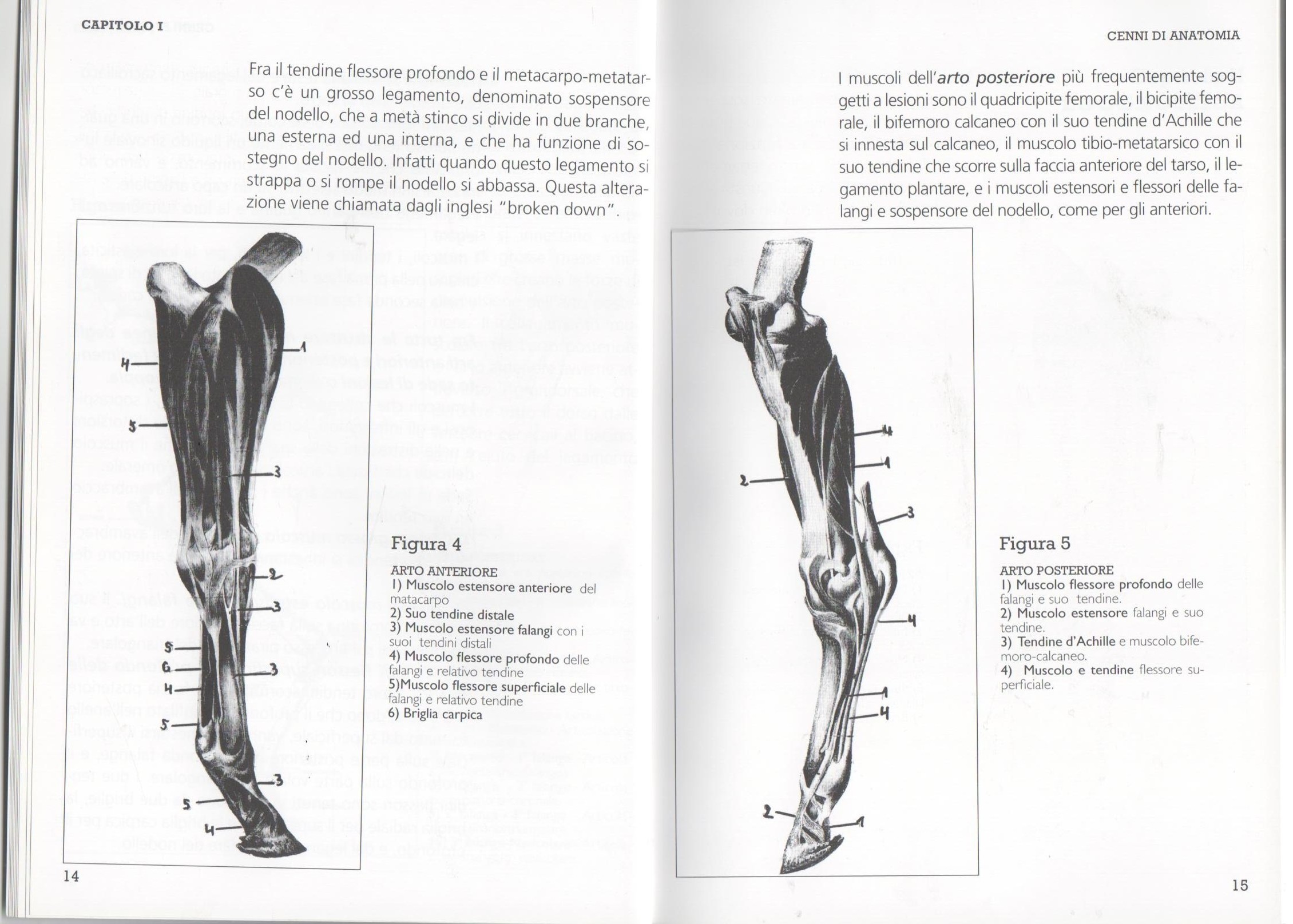
What’s more, particularly prone to injury are also the forearm muscles and their respective tendons such as the short and thick extensor muscle, of which its tendons are inserted on the front of the carpus. As for the superficial and deep muscles of the phalanges, their tendons flow on the rear face of the limb.
The two flexor tendons are held in place by two bridles, the radial for the surface and the carpal for the deep, and by the annular ligament of the fetlock. Between the deep flexor tendon and the metacarpal-metatarsal, there is a major ligament, called the suspensory fetlock, which in mid-shin splits into two branches, one outside and one inside, which is designed to support the fetlock.
In fact, when it tears or breaks, this alteration is known as “broken down“. Among the hind limb muscles, the more prone to injuries are the quadriceps and the hamstring, the Achilles tendon that inserts on the calcaneus, the tibia-metatarsal muscle with its tendon that runs on the front face, the plantar ligament and the extensor and flexor muscles of phalanges and the suspensory fetlock.
The following was adapted from the book “La zoppia del cavallo, conoscere per capire / the lameness of the horse, learn to understand“ by Dr. Vittorio Meschia, edition by Horse s.r.l. editore.


.png)
