The Importance of Equine Odontostomatological Care: Radiography and Advanced Diagnosis
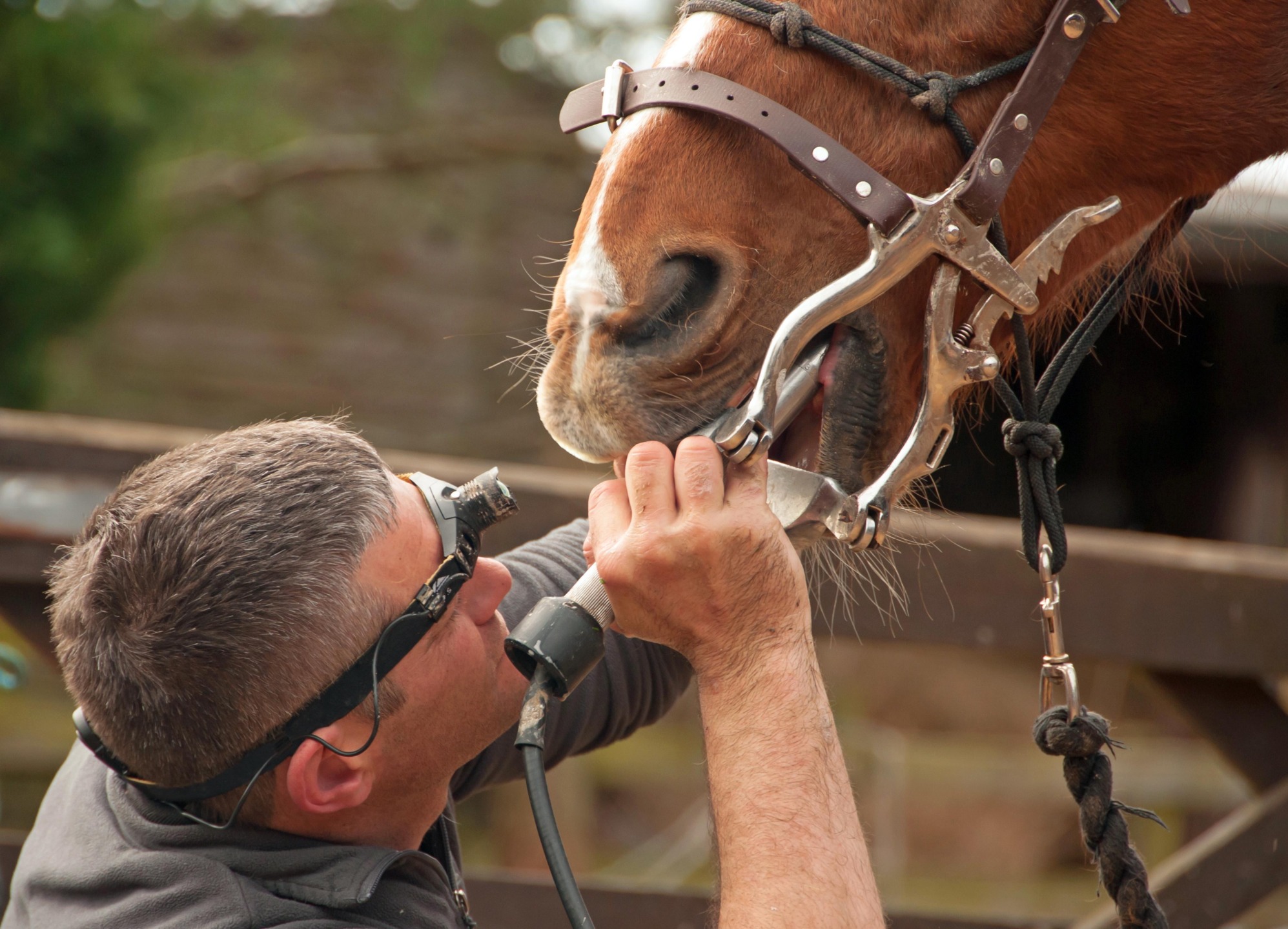
Understanding Equine Oral Health to Prevent and Treat Diseases
Dental health is a fundamental element of a horse’s overall well-being. Undiagnosed dental issues can negatively affect performance, behavior, and the general health of the animal. In this context, diagnostic imaging—particularly radiography and computed tomography (CT)—has revolutionized equine veterinary medicine, enabling accurate and timely assessment of dental and sinus pathologies.
The Evolution of Equine Dental Imaging
Radiography is the first-line tool used to investigate dental or sinus abnormalities that are already suspected during a clinical examination. It allows visualization of bony and dental structures such as the lamina dura, alveolar bone, mandibular and maxillary arches, nasal cavities, and paranasal sinuses.
However, for a more precise diagnosis and for viewing complex areas without overlapping structures, computed tomography now represents the most advanced diagnostic standard. The sectional nature of CT provides significantly higher resolution and a detailed identification of pathologies, especially in deep areas of the skull.
Dental Radiography: Indications and Techniques
The horse’s head, due to the combined presence of air and bone, offers good radiographic contrast. Nevertheless, its anatomical complexity can make it difficult to detect subtle lesions. It is therefore essential to perform the examination with the horse sedated to prevent movement.
The main indications for dental radiography include:
- Cranial trauma
- Supernumerary teeth
- Dental fractures
- Apical abscesses
- Deep periodontal disease
- Retained deciduous teeth
- Facial swelling or unilateral nasal discharge
Radiographic Techniques for Incisors and Canines
Thanks to their position, incisors and canines are relatively easy to radiograph. Intraoral placement of the radiographic cassette between the incisors allows for good visualization, with beam angulation calculated according to the bisecting angle technique. Canines can also be visualized using similar techniques, with lateral angulations of about 45°.

Radiography of Maxillary and Mandibular Teeth
Extraoral radiographs with the mouth closed are commonly used to study cheek teeth and sinuses. Standard views include:
- Lateral (LAT): useful for evaluating sinuses and occlusion.
- Open-mouth view: reduces overlap, improves crown visualization.
- Oblique dorsoventral: enables examination of upper molar roots and maxillary sinuses.
- Dorsoventral (DV): good for visualizing central skull structures.
For mandibular teeth, the presence of the masseter muscle and the narrow space between the mandibular rami make imaging more challenging. In such cases, a ventrodorsally oblique projection is preferred to improve root visualization.
Intraoral Radiography of Molars
Currently, intraoral radiography in horses mostly uses portable CR or DR panels measuring 24×30 cm, which still have several limitations. Recent solutions such as larger intraoral sensors and PSP readers allow for high-definition images, reducing overlap and improving diagnostic accuracy.
Effective sedation is crucial to avoid tongue movement and chewing. This type of examination allows for analysis of:
- Height of alveolar bone
- Thickness of the periodontal ligament
- Density of the lamina dura
- Presence of dental fragments
Radiographic Interpretation: What to Look For
Knowledge of radiographic anatomy is essential for correct interpretation. Key features include:
- Dental apices: the terminal part of the root
- Lamina dura: compact bone surrounding the root
- Periodontal ligament: thin radiolucent line between tooth and bone
- Alveolar crest: bony projection between adjacent teeth
Radiographic signs of disease include:
- Periapical lucency (sign of apical abscess)
- Blunted or deformed apices (sign of chronic inflammation)
- Irregular thickening of the lamina dura
- Widening of the periodontal space
- Signs of maxillary sinusitis (diffuse opacity and fluid levels)
- Alveolar bone resorption (sign of advanced periodontitis)
Radiography is an essential diagnostic tool for monitoring and treating oral and sinus pathologies in horses. The combination of intraoral and extraoral techniques, along with the growing availability of computed tomography, now allows for precise and early diagnosis, with positive impacts on the animal’s health, comfort, and performance.
Investing in Equine Odontostomatological Care means not only preventing silent diseases, but also ensuring a longer, healthier, and more performant life for our horses.
© Rights Reserved.


.png)


